Wednesday, August 31, 2011

Week 8


In the eighth week, my adviser asked me to stay at OIMB for one extra week, in order to polish up the website. I happily agreed, so I'll be here for a total of nine weeks. The website definitely needs a good deal of work so I am happy to put in more hours on it. Though to be honest, photo processing is exhausting work!

Most of this week was spent preparing for final presentations, which took place on Friday. I spent most of Tuesday reviewing notes on good ways to structure presentations. I gave a few practice presentations to my friends and felt ready to present on Friday. The presentations took place in a classroom at the Oregon Institute of Marine Biology. Unfortunately the Hatfield Marine Science Center interns did not come down to Charleston for the day, but we did get the chance to see their presentations via Skype.

During my presentation, I spoke briefly about my future plans for the website (akiko-invertebrates.weebly.com), which I made to house the identification key which is my summer project. With my extra time at OIMB, I hope to label the basic anatomy of the juvenile settlers, and post a few photos of what each settler looks like as an adult colony. The website needs a lot of editing, and I have received many helpful comments about how to improve it. I hope that I have enough time to apply them all!


This is me looking through the dissecting microscope, which I used for the bulk of my work. Every day, for four weeks or so, I looked through this microscope to take pictures of the settling organisms.


A friend of mine took this photo of me and some ascidians. The large orange blobs are Distaplia occidentalis colonies, growing on a piece of netting that has been submerged for several months.

Wednesday, August 17, 2011

Week 6

The weeks have flown by! I have been hard at work. During the sixth week, I finally stopped taking daily photos of the settlers. It's fun to do work under the microscope, but hard when you're doing it every day! I compiled all of the photos I took (over 2,000!) and began sorting through them in order to find the highest quality ones for each species. Once I had chosen the nicest photographs, I began the long process of modifying them with Adobe Photoshop. Since I am new to both microscopy and photography, most of the pictures that I took were blurry, too dark, too bright or off-colored. Additionally, many of the organisms that I photographed are translucent, making them even harder to see on a picture. With Adobe Photoshop, I can adjust the photo to make the image clearer and more representative of the actual organism.

In addition to image quality modifications, I can also use Photoshop to create composites of multiple images. This is a very useful way to deal with the limitations of microscopy. With microscopes, the focal plane-- that is, how much of the object is in focus-- is very thin. This means that at any one time, only some parts of the 3D organism will be in focus. In any one picture, only a portion of the organism will be in focus and the rest of the organism will be blurry, as shown below. However, with Photoshop, I can slice out the parts from each picture that are in focus, and combine them together to make a composite image in which most of the organism appears to be in focus.

Here, the base of the organism is in focus.


Here, the top part of the organism is in focus.


This picture is a composite that I have made using Photoshop, by combining the two above images.


Photo processing takes a good deal of time to do, and I spent many hours going through the photos in order to get them cleaned up and presentable. In addition to that, I also began to learn how to make a website on using the "Weebly" website creation page. Thankfully, the interface on the Weebly page is very user-friendly. I am still ironing out the finer points of the web page but so far it is looking good! It is still under construction, but is viewable at: akiko-invertebrates.weebly.com .

Monday, August 8, 2011

Week 5

In the fifth week of the internship, all of the OIMB interns in Charleston and the Hatfield Marine Science Center interns from Newport got the chance to come together and show off our projects. The COSEE PRIME program makes a huge effort to support communication and collaboration among scientists, which is such a great initiative. Having spent many hours in the lab, I can see how a researcher could become so involved in their work that they don't get the chance to see what other scientists are doing in similar fields. However, collaboration is so important, because the results that you get under a microscope could heavily influence the work of another researcher doing a similar project.

On Wednesday, Coty and Drew came down to Charleston with their adviser Itchung. I showed them all my project and let them watch the little ascidians pump water into themselves. Every once in a while, the organisms will contract their branchial baskets and close their siphons as if they are coughing or exhaling. From what I've read, this is to keep large particles from entering their incurrent siphons. This reaction is touch-sensitive, so sometimes I will tickle the ascidian with a single hair (that's how small and sensitive they are) and watch them contract. Later on that afternoon, we all went out with Professor Jan Hodder to see some marine mammal action at Cape Arago. Seals and sea lions sunbathed on the rocks, and a few gray whales broke the surface every few minutes.

On Thursday, Karyn, Jess, and I with our adviser, Coral, went up to Newport to visit with the interns there and see their projects. This was the first day in over 3 weeks that I took a full day off and didn't look at my petri dishes! It was nice to have a little break and it gave me time to reflect on my project progression and to write in my research notebook. We got to stomp around in the mud with Drew, and visit Coty's bird-watching location. It was a nice trip, but good to come back home!

Playing around in the mud was fun. I got to use a huge siphon to suck shrimp out of the mud. We saw ghost shrimp and mud shrimp.


We had a blast looking at marine mammals at Cape Arago!

Wednesday, July 27, 2011

Week Four

Hey All, Akiko here.

My goodness the weeks are passing by so quickly! During the fourth week, I watched my six new plates get colonized by a variety of settlers. The plates are fouling quickly, which is good because it means that a lot of organisms are settling down on it, but is also difficult because algae and particles have clouded the petri dish and make it hard to clearly see the organism in photos.
I decided to make a little side-experiment out of the redeployment. I actually deployed 12 plates with velcro on the sides instead of underneath. Of the 12 plates, six of them have abrasion on the top side (I scrubbed them with sandpaper to make the surface rough) while six of them are simply smooth plastic. I did this because supposedly settlers prefer to settle on roughened surfaces, and I figured that I could keep track of the rate of settlement on the clear plates versus the smooth plates. I'll count the number of settlers on each plates at some future time during the experiment and see whether the settlers really prefer rough over smooth.

For my main experiment, every day is the same procedure: get the petri dishes out of the water, look at each of them under the dissecting microscope, take photos of specific organisms, and keep track of new species that arrive on my plate. It sounds easy, but the entire process takes about 3-4 hours, and I do it every day (even the weekends). I am trying hard to be a good scientist, even if it means sacrificing my days off. On the bright side, once I start looking at the plates I'm often so absorbed in observing the changes that go on from day to day that the time goes by faster than it would otherwise.

Here are some of the new organisms that have collected on my plates. I haven't had time to adjust the images or put in scale bars, but suffice to say that they are really tiny!

This is probably Schizoporella japonica, a bryozoan. Bryozoans are little animals that settle onto surfaces and reproduce asexually to form larger colonies. This kind of bryozoan is an encrusting bryozoan, meaning that it will continue to spread over the surface in a hard thin layer, like dried paint. When this bryozoan gets bigger, it will begin to reproduce assexually to form a colony.


The spiral shell in this photo belongs to what I believe is a serpulid polychaete worm. You can see little feeding structures sticking out of the mouth of the shell. That is the worm. With these guys, I have to wait for them to peek out of their shells before photographing them.

Friday, July 15, 2011

Trial and Error

((these posts are taken straight from the blog that I have to write for my internship, so they're not quite as personal))

In the third week of my internship, I spent a lot of time focused on my research project. I spent a lot of time diligently following, photographing, and observing the little ascidians that settled on my plate. I used velcro on the backs of my petri dishes to attach them to a large plate hanging in the bay. However, this velcro created a problem in that it obscured the light from the microscopes when trying to take pictures of the ascidians. Furthermore, I found that my original intention of setting out 20 petri dishes was too ambitious: I quickly found myself inundated with work trying to photograph 20+ organisms each day!

I decided that I needed to rethink and redo some parts of my project. With the guidance of my adviser, Professor Richard Emlet, I made new petri dishes that had velcro on the sides of the plates rather than underneath them. That way, the velcro wouldn't distort the light. I also decided to downsize the number of petri dishes deployed. Now I have six clear petri dishes to keep track of instead of twenty. I also decided not to take pictures of all of the organisms each day, but to rotate through them.

On the up side, I am learning something new every day. Each day I dedicate a lot of time (around 3 hours) to observing and photographing the tiny organisms under the microscope, and then I put in a few hours to tidying up the images with photoshop. I'm learning how to recognize settlers of different species too! Here are some that are easy to identify.


Do you recognize this one? This is Botrylloides violaceus, the pictures that I posted on my last blog entry. They are generally easy to recognize because of their huge sunburst-like ampullae and their red, orange or purple hue. They are also relatively large compared to the other early settlers that I see under the microscope.


These next two images are of a different species of colonial ascidian. This is called Distaplia occidentalis. In these photos, you can see three or four little ampullae that look like legs of a tripod in contact with the surface of the petri dish. You can also see a large cylindrical shaped structure that looks like a mesh tube. That is called the branchial basket, and it's connected to the organism's
incurrent or branchial siphon- the nozzle that sucks water into the organism.


Yet another organism that fouls my plates: the sponge. Unfortunately, I don't know the scientific name of this one. However, I do get a lot of sponges on my plates and I take pictures of them, even though they aren't as charismatic as the colonial ascidians.

Wednesday, July 6, 2011

Project update

So I spent most of the first week researching and designing my project, and during the second week I got to finally start implementing it. After a lot of planning I figured out a way to catch some pictures of microscopic settlers as they mature.

First I sanded the surface of a few petri dishes in order to make an abrasive surface. Supposedly, invertebrate settlers find the roughened surface more appealing to attach to. Then I attached velcro strips to the back of each petri dish with glue. I put longer strips of velcro on a large rectangular plate. This large rectangular plate has four holes drilled into it and rope tied to it, such that it can hang from a dock and be suspended in the water column. After lots of preparation, I attached my petri dishes to the backing plate and lowered them into the water for a few days, checking them every day.

I kept track of individual organisms by making a small grid that can fit over each petri dish, so that I can track their location and confirm that it's the same organism. It's very hard to keep track of them when they are so small! Every day, I took a photograph of each invertebrate settler. I have found that it is difficult to get good images when you're taking a picture through a microscope, but with some lighting adjustments and photoshop help, I hope to get images that could be used for identification.


These images are all of the same organism. I'm pretty sure that the scientific name is Botrylloides violaceus. I have had quite a number of these settle on my petri dishes. They are easy to identify because of their ampullae: the sunburst-like ring around them.

Saturday, July 2, 2011

Beach Day- Oregon Coast style

I spent my Saturday hanging out with the other OIMB kids at the beach! A bunch of us crammed into a car and drove over. This beach is different from the beaches at home. I'm used to Manele/Hulopo'e bay: where the waves break one by one on the sand and the ocean floor drops off to 6 feet or so and then gets deeper from there. Here, the sand slopes down into the water very gradually, such that you can walk out away from shore for at least 20 yards and still be able to stand. However, there are constant waves breaking over you and wicked cross current. Not to mention, the water is around fifty degrees Fahrenheit! For those of you who have never experienced water like that, it means that as soon as you walk in the first sensation you feel is cold, and the second is a flood of pain. It takes about ten minutes for each part of your body to slowly become numb, but until it does, it is pretty excruciating. Last week, I was proud of myself for staying in the water for around 35 minutes without a wet suit. This week however, there was a spare wetsuit so I put it on and went swimming with a few other students. It was fun to be in the water again, though the white wash was battering my face and body with each wave. In fifty degree water it is physically painful to dunk your head in, so duck-diving under the waves was a little difficult.

After my swim, people played football and beach ultimate frisbee. I wanted to play ultimate badly, but I have a terrible blister on my foot that has refused to heal. Just walking on the sand was painful, so I sat out of the game and laid in the sun. Really, not too bad of an option in itself.

So far my experience here has been wonderful. The students are inclusive, accepting, fun-loving and enthusiastic. I'm so glad that they have all made it easy to have friends here. Hanging out with all of them really defines the experience as a positive one for me. The amount of joy and satisfaction in my life relies so heavily on attitudes of people around me, and I am very thankful for all of the positivity and support that I've gotten from the OIMB students.

Friday, July 1, 2011

Some Pictures

The two mushroom like blobs on the left are Distaplia occidentalis, colonial ascidians. The pink colored blob on the right is a sponge, Haliclona sp.


This is Didemnum vexillum the invasive species that has been spreading around the globe. It is also a colonial ascidian, meaning that this blob is made up of thousands of tiny individuals called zooids.


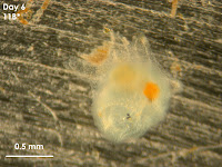
This is from my first day of collecting data! I took this picture of a Botrylloides settler who has just metamorphosed from a free swimming larva to a settled organism. The little sun burst like arms are called ampullae. The main "body" where most of the dark pigment is, is the first zooid (individual) to settle down and is called the oozooid. The oozooid then begins asexual reproduction by budding off new copies of itself, called blastozooids. You can see that this specimen is just starting to make blastozooids- they are the transparent blobs coming off of the top and bottom of the oozoid.

Thursday, June 30, 2011

Summer 2011 at OIMB

Yet another attempt at keeping an updated blog :)
I am at the Oregon Institute of Marine Biology, where I will be spending my summer. I am required to keep a blog, and the official blog is at http://coseeppprime.blogspot.com/ However, as that is the official blog, I'm going to repost them here on my blog and also embellish those posts with my own personal feelings as well.
Anyhow, here's my first post. Pictures will be added soon :)

My name is Akiko Onuma, and I am lucky enough to be one of the COSEE Pacific Partnership PRIME interns. About a week ago, I wrapped up my finals and projects at Portland Community College and moved out here to the Oregon Institute of Marine Biology in Charleston, Oregon. It’s my fourth day here and so far I’ve had a pretty awesome time!

On my first day of the program, I met with my mentor, Professor Richard Emlet, to get an idea of the purpose of my project. He told me about the many different invertebrates living in the two boat basins in Charleston. A few of them include colonial invertebrates, where hundreds of tiny individual organisms make up a larger colony. The organisms that I will be studying are called fouling species, meaning that they attach themselves to hard surfaces underwater. Most of them look like slime growing on rocks or boat hulls, which is how they got their name "fouling organisms". My project will be to make a photographic timeline/identification key that will look at a variety of these marine invertebrates from when they are very small to when they are much bigger. Most of these organisms go through a larval period, meaning that they start off as a microscopic swimmer floating through the water. Soon, they find a hard surface and settle down on it, undergoing metamorphoses in order to attach themselves to the substrate. Then, they start to “grow” by asexual reproduction, which basically means making more of yourself in order to start a colony. It is the colony that is visible to the naked eye, and identifiable. So my job is to take pictures of these species from the time when they first settle (attach themselves to the surface of my petri dishes) until they are big enough to be identified.

Harbors and marinas are often hotbeds for introduced and invasive species because boats will pick up and distribute these hitch-hiking species wherever they dock, much like the marks left by a child playing with a jar of glitter. One particularly important introduced species is called Didemnum vexillum. This colonial organism, which looks like pale orange slime growing on the rocks, is an invasive species. Scientists believe that this organism originally came from Japan, but it is showing up in docks all over the world. Didemnum has documented presence in Japan, New Zealand, France, Ireland, California, New Hampshire, and now recently has started settling in Oregon. Furthermore, once introduced, Diademnum grows rapidly and can cover large areas in a short period of time, out competing other native species for space and resources.